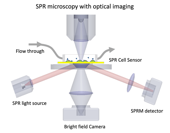
SPR microscopy integrates optical imaging and SPR technology into one system. It provides high-quality data on binding kinetics and affinity of cell membrane proteins and spatial visualization of cellular structures. Real-time interactions of the drug and membrane protein can be measured in its native state without needing to extract proteins from the cell. Because of its unique measurement of cell membrane proteins on the cell, SPR microscopy has become a powerful tool that provides binding information about various drug compounds and drug efficacy. Antibody-interaction studies are also used to investigate disease pathways and targets for potential drug therapies, and much more.
In addition, the system measures nanometer scale binding activities needed for the study of bacteria and virus interactions and the development of new methods for nanoparticle drug delivery.
- Technical Note 105: Introduction of SPR Microscopy
- Application Note 122: Surface Plasmon Resonance Microscopy for Multiple and Single Cell Membrane Binding Kinetics Studies
- Application Note 124: Quantifying Molecular binding to Membrane Proteins on Individual Cells with Surface Plasmon Resonance Microscopy
- Application Note 125: Lectin-Glycoprotein Interactions with SPR Microscopy
- Application Note 126: GPCR Binding Assays with SPR Microscopy
- Application Note 127: Quantifying Antibody Binding to Membrane Proteins on Single Cells with SPR Microscopy
- Application Note 128: SPR Microscopy for Acid-Sensing Ion Channels
- Application Note 129: AZ1395 small molecule targeting GPR39
- Application Note 130: Measuring molecular binding kinetics with SPRM Impedance
- Application Note 131: SPR Microscopy for Live Suspension Cells
- Application Note 132: SR142948 Antagonist Binding to NTS-1 Receptor Using SPR Microscopy