Neurotensin receptor belongs to the family of G protein-coupled receptors (GPCR), which has 424 amino acids with 7 putative transmembrane domains.(1) Neurotesin receptor 1 (NTS-1) mediates multiple functions of neurotensin, such as hypotension, hyperglycemia, hypothermia, antinociception, and regulation of intestinal motility and secretion.(2) Despite the importance, a suitable assay that can directly measure GPCR-drug interactions has been challenging, due to issues associated with allosteric modulation, probe dependence and functional selectivity.(3,4)
In this application note, we present a study of the binding of S142948A, a non-peptidic small molecule neurotensin receptor antagonist (685.9 Da) to NTS-1 using SPR microscopy (SPRm 200) without any special protocols. SPR microscopy is a label-free technology that measures binding kinetics of membrane proteins without extracting the proteins from the cell membranes, which helps preserve the native conformations of the membrane proteins.(5)
Endogenously expressing NTS1 cells (MCF7 breast cancer) were cultured on a collagen coated SPRm sensor chip. Titration of SR142948 antagonist up to 50 nM (dilution factor 2) were flown over the sensor chip. Using ImageSPRTM software, hundreds of ROIs (Region of Interest) were analyzed to provide statistical analysis of binding affinity and kinetic constants of the individual cells.
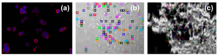
FIG. 1 (a) ICC (immunocytochemistry) validation of NTS1 expression in MCF7 (Hoechst stained). (b) Bright field image of MCF7 cells on the SPRm chip with selected ROIs. (c) Simultaneously recorded SPRm image of S142948 binding to NTS1 receptor.
The measured key binding parameters include: KD= 2.03 nM with 95% CI 1.21 – 3.38, ka= 2.19×10⁶ M-1 s-1 (CI 1.76 – 2.72) and kd= 4.74×10⁻³ s-1 (CI 3.38 – 6.66). The SPRm affinity data is close to the functional assay data obtained for the wild-type NTS1 receptor (IC50= 8.4 +/- 0.9 nM).(6) KD extracted from the traditional SPR using a biophysically stabilized variant of NTS1 (NTS1-H4 receptor) is 0.4 nM.(7)
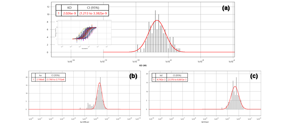
FIG. 2 (a) Histogram of measured KD with dose response curves of all ROIs (b) ka and (c) kd.
In conclusion, SPRm 200 integrates the biophysical analysis of binding affinity and kinetics of drug targets and the viewing of cells in a physiological setting. This technology delivers binding kinetic data for important drug targets but difficult to assess with the traditional technologies.
We thank Dr. Jamie Ware and Dr. Scott Pollack from Sygnature Discovery for the samples, data and valuable insights.
Author: Nguyen Ly | Biosensing Instrument | Published Jan 4, 2025
DOWNLOAD PDF
Download a PDF of Application Note 132: SR142948 Antagonist Binding to NTS-1 Receptor Using SPR Microscopy
- Tanaka, K. et al, Neuron. 1990 Jun, 4(6): 847-54
- https://www.ncbi.nlm.nih.gov/gene/4923
- Navratilova, I. et al, ACS Med. Chem. Lett. 2011, 2,7, 549-554
- Navratilova, I. et al, Anal. Biochem. 2006, 355(1):132-9
- Wang, W. et al, Nat. Chem. 2012, 4, 846-853
- Egloff, P. et al, Proc Natl Acad Sci USA 2014, 111(6): E655-62
- Huber, S. et al, PLoS ONE 2017, 12(5) e0175842