The cell-based Surface Plasmon Resonance Microscopy (SPRM) system, integrates optical microscopy and SPR, is a powerful technique for measuring binding activities of membrane proteins in vitro. It allows the simultaneous measurement of phenotypical changes of the sample via bright field and binding strength and kinetics via SPR.
Click here to download a PDF datasheet.
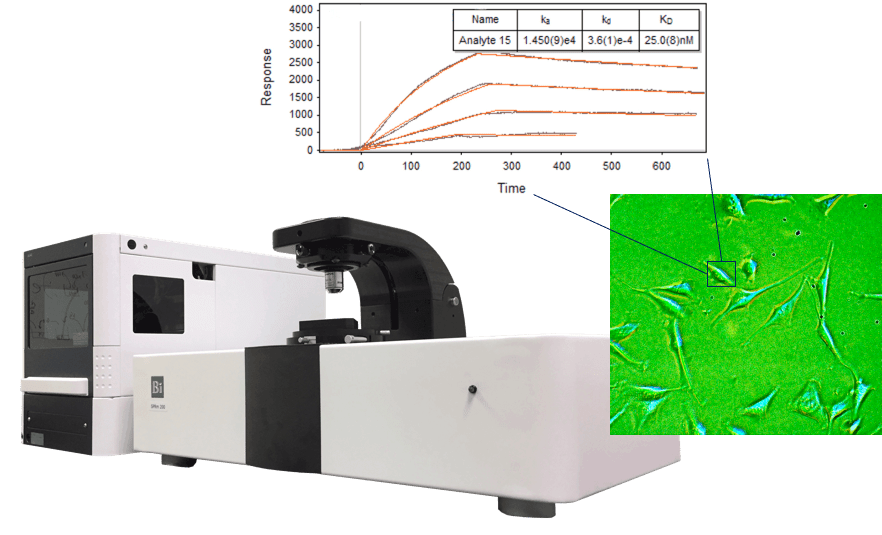
- Integrated optical microscopy with SPR Bright-field and SPR microscopies in one instrument. Large field of view with high resolution optics to view single or multiple cells.
- In vitro and label-free binding activities mapping Provides SPR sensorgrams and binding activity map of individual cells as well as the binding kinetic constants (ka, kd, KD).
- Nanometer scaled binding response of virus, bacteria and nanoparticles Nanomotion detection study of bacteria or virus metabolic activity or binding activities of nanoparticles for drug delivery designs.
- Technical note #106 “SPR Microscopy vs Radioligand Binding Analysis” TechNote106
- Technical note #105 “Introduction of SPR Microscopy” TechNote105
- See SPR Microscopy AppNotes
- K Syal, R Iriya, Y Yang, H Yu, S Wang, S Haydel, HY Chen, NJ Tao, “Antimicrobial Susceptibility Test with Plasmonic Imaging and Tracking of Single Bacterial Motions on Nanometer Scale“, ACS Nano, 10, 845-852, 2016
- F Zhang, S Wang, L Yin, Y Yang, Y Guan, W Wang, H Xu, N.J. Tao, “Quantification of Epidermal Growth Factor Receptor Expression Level and Binding Kinetics on Cell Surfaces by Surface Plasmon Resonance Imaging“, Analytical Chemistry, 87(19), 9960-9965, 2015
- L Yin, W Wang, S Wang, F Zhang, S Zhang, N.J. Tao, “Measuring Binding Kinetics of Antibody-Conjugated Gold Nanoparticles with Intact Cells”, Small, 2015
- W Wang, L Yin, L G-M, S Wang, X Yu, S Eaton, S Zhang, H Chen, J LaBaer, N.J. Tao, “In situ drug-receptor binding kinetics in single cells: a quantitative label-free study of anti-tumor drug resistance”, Scientific reports, 4, 1-7, 2014
- W Wang, Y Yang, S Wang, V Nagaraj, Q Liu, J Wu and N.J. Tao, “Label-free measuring and mapping of binding kinetics of membrane proteins in single living cells“, Nature Chemistry, 4, 846-853, 2012
- W Wang, K Foley, X Shan, S Wang, S Eaton, V Nagaraj, P Wiktor, U Patel and N.J. Tao, “Single cells and intracellular processes studied by a plasmonic-based electrochemical impedance microscopy”, Nature Chemistry, 3, 249-255, 2011