
Staphylococcus epidermidis is a common skin bacterium and a major contributor to healthcare-associated infections because it colonizes medical devices, forms biofilms, and frequently exhibits antibiotic resistance.1 Antimicrobial screening usually measures growth inhibition, but these endpoint assays do not show whether an agent binds directly to the bacterial surface, how rapidly it associates, how much remains after washing, or whether all bacteria respond similarly.2
Surface plasmon resonance microscopy (SPRM) addresses these questions by combining label-free, real-time detection with microscopic localization. In this study, live stationary-phase S. epidermidis cells were immobilized directly on a chemically modified gold sensor. The sensor was cleaned with water and ethanol, annealed with a hydrogen flame, incubated overnight with 1-dodecanethiol (DDT), and treated with 1% 3-aminopropyltriethoxysilane (APTES). A 2 µL aliquot of the S. epidermidis cells suspension was then drop-cast onto the modified sensor and air-dried to establish stable attachment. The immobilized bacteria were exposed to phospholipid liposomes containing the antimicrobial host-defense lipid cholesteryl linoleate (CL-PL), while phospholipid-only liposomes (PL) served as carrier control. As the immobilization procedure involved chemical surface modification, bacterial deposition, air drying, chip mounting, and continuous buffer flow, it was important to confirm that the experimental workflow itself did not eliminate bacterial viability. Tetrazolium agar testing performed immediately after bacterial deposition and following a complete buffer only SPRM run confirmed that the immobilized bacteria remained metabolically active. Although this assay did not assess full bacterial functionality, it demonstrated that the bacteria remained viable throughout the SPRM procedure in the absence of antimicrobial treatment.
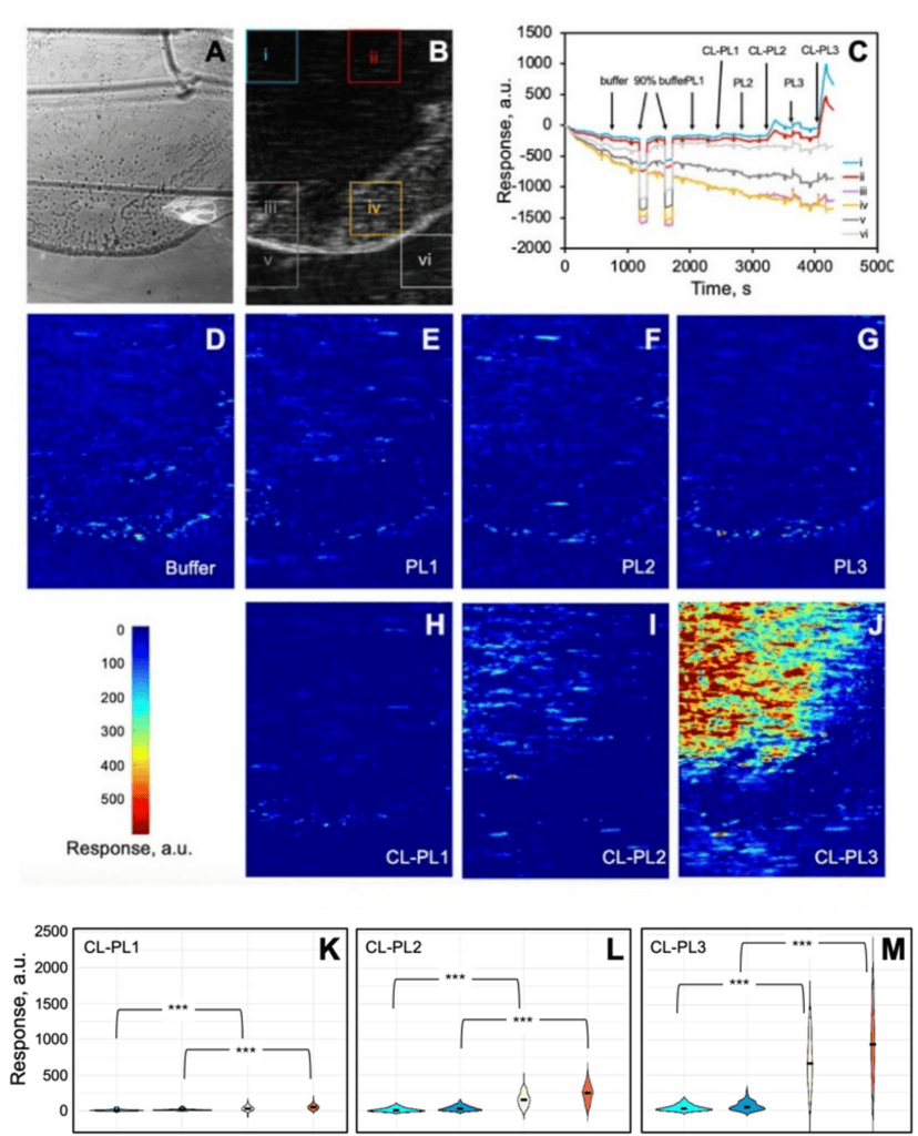
Figure 1: Representative surface plasmon resonance microscopy (SPRM) results showing liposome interactions with immobilized bacteria. (A) Bright-field image and (B) corresponding SPR image highlighting the boundary of the bacterial deposition region. (C) Real-time SPRM signals extracted from six regions of interest (ROIs) indicated in (B). (D-J) SPRM images at the end of each injection after subtracting the corresponding first frame. All images are 600 μm × 450 μm in size. SPR image stacks were converted to TIFF stacks using ImageAnalysis (Biosensing Instruments, Tempe, AZ) and then converted to image sequences using ImageJ for further analysis. (K-M) Quantitative comparison of the intensity changes among injections from H to J shown in violin plots. The width of the violins indicates the population density at a given response unit, while the black bar in the center represents the median response. ∗∗∗p < 0.001. Light blue (background) and white (bacteria) plots represent the “sustained” signal changes by the end of each injection, while dark blue (background) and orange (bacteria) plots represents the “temporary” signal changes during the exposure stage.
SPRM imaging distinguished bacteria covered regions from bacteria-free background regions on the same sensor surface, while sensorgrams from selected regions recorded the binding response in real time (Figure 1A-C). CL-PL were tested at CL concentrations of 1.54, 7.70, and 38.5 µM and compared with PL. PL produced minimal binding responses, whereas CL-PL showed clear association and dissociation profiles that increased with CL concentration (Figure 1C, D-J). The signal that remained after buffer washing indicated that CL-containing liposomes accumulated on and remained associated with the bacterial surface.
SPRM response maps showed that CL-PL accumulation was concentration dependent and spatially heterogeneous across the immobilized bacterial population (Figure 1H–J). The lowest CL-PL concentration produced only a weak and localized response (Figure 1H), whereas the number and intensity of responsive regions increased at the intermediate and highest concentrations (Figure 1I–J), with the strongest hotspots observed at the highest concentration. In contrast, buffer and PL-only liposomes produced minimal sustained responses across the three concentrations (Figure 1E–G), indicating that the observed accumulation was primarily associated with the cholesteryl linoleate component rather than the phospholipid carrier. ROI based analysis showed that CL-PL injections generated higher “Temporary” and “Sustained” values and established a dose dependence at the bacteria regions than at the background regions (Figure 1K-M). The bacteria were collected after approximately 20 hours of growth and were therefore in the stationary phase, during which the population may contain actively metabolizing, metabolically inactive, stressed, damaged, and nonviable cells with differences in gene expression and surface composition. Cell-to-cell differences within the stationary phase bacterial population including variations in metabolic state, viability, gene expression, and surface composition could affect the availability of surface binding sites and the extent of CL-PL association or retention. Although the study did not directly link individual hotspots to specific bacterial phenotypes, the findings demonstrate that SPRM can detect spatial heterogeneity across a bacterial population. This capability could be valuable for investigating bacterial persistence, antimicrobial tolerance, and other heterogeneous responses.
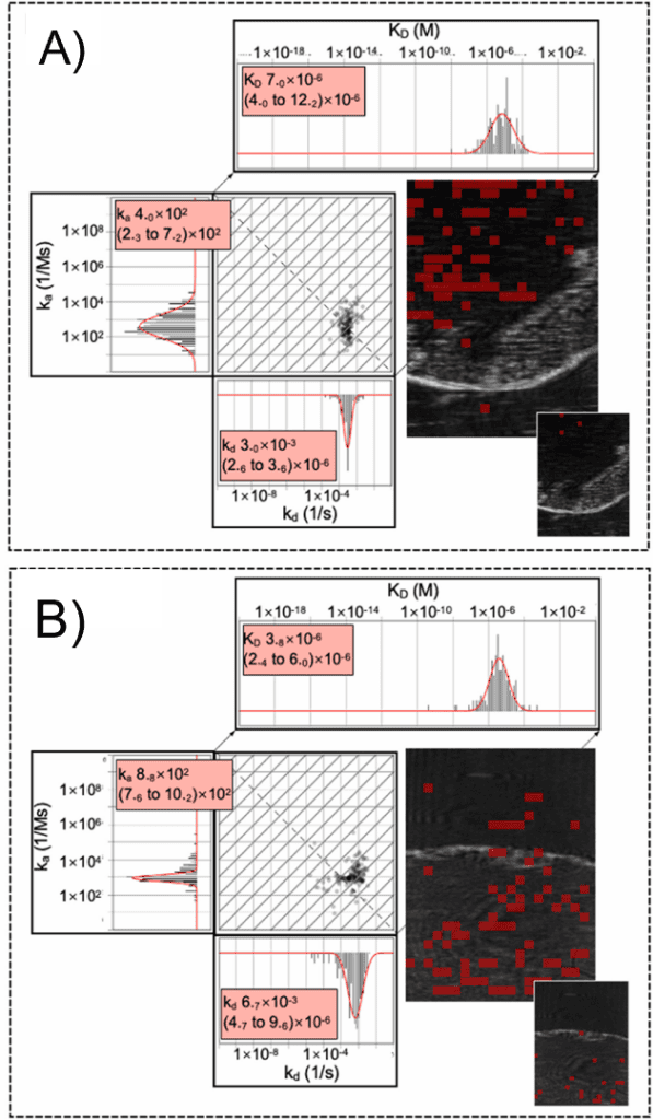
Figure 2. Spatially resolved kinetic analysis of CL-PL liposome binding to S. epidermidis. (A, B) Isoaffinity plots of the association rate constant (ka) versus the dissociation rate constant (kd) from two independent experimental runs, showing the distribution and heterogeneity of the bacteria–CL-PL interactions. The accompanying histograms and Gaussian fits show the distributions of ka, kd, and the equilibrium dissociation constant (KD), with mean values and 95% confidence intervals. The two runs yielded KD values of approximately 7.0 and 3.8 µM. The corresponding SPRM images show the spatial distribution of ROIs that met the kinetic-fitting quality criteria. Larger SPR images represent CL-PL injections, smaller SPR images (inset)represent PL-only controls, and red boxes indicate ROIs with valid kinetic fits.
Kinetic fitting localized most valid CL-PL interactions to bacteria-covered regions. Despite differences in bacterial coverage and absolute response, the two experimental runs yielded equilibrium dissociation constants of approximately 7.0 and 3.8 µM (Figure 2). In addition to quantifying binding kinetics, SPRM mapped the spatial distribution of these interactions across the bacterial sample. This approach identified where binding occurred, enabled local background correction, distinguished temporary responses during sample exposure from sustained liposome retention after washing, and revealed regional differences in CL-PL binding. These capabilities make SPRM well suited for comparing antimicrobial formulations, bacterial strains, and growth conditions, as well as for investigating heterogeneous responses within live bacterial populations.
Materials is a summary of Chan et al Biosensors and Bioelectronics: X (2026): 100792.
Author: Nguyen Ly, and Miyuki Thirumurthy | Biosensing Instrument | Published November 11th, 2026
DOWNLOAD PDF
Download a PDF of Application Note 184: Real Time Spatial Analysis of Antimicrobial Binding to Live Bacteria by SPR Microscopy
- Burke et al, Virulence 15.1 (2024): 2359483.
- Kadeřábková et al, Antimicrobials and Resistance 2.1 (2024): 37.
- Chan et al, Biosensors and Bioelectronics: X (2026): 100792.