
Neuronal cell-based studies are important in drug discovery because many neurological, neurodegenerative, pain, and neurodevelopmental diseases depend on cell specific receptor signaling, neurite formation, synaptic biology, survival pathways, and toxic responses that cannot be fully captured in traditional biochemical assays alone.1 Neuronal cell models help place surface binding measurements in a more physiologically relevant cellular context than purified protein assays alone.
In this application, PC12 (adrenal gland tumor) cells are differentiated into neuronal cells on an SPRM chip using nerve growth factor (NGF), enabling label-free analysis of neuronal morphology and surface binding behavior in a format compatible with kinetic measurements. PC12 cells are a useful model system because NGF stimulation activates TrkA signaling and drives a well-characterized transition from proliferative chromaffin-like cells to neuronal cells with neurite outgrowth, making the system practical for studying neuronal differentiation and receptor-mediated responses.2
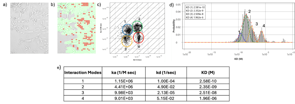
Figure 1: Kinetic analysis reveals four distinct neuronal binding populations:(a) brightfield imaging shows immobilized neuronal cells before antibody injection, and (b) the SPR binding map shows localized anti-TrkA binding events across the neuronal cell surface,(c) ka/kd isoaffinity distribution of antibody binding identified four heterogeneous affinity populations, with four different KD values supporting heterogeneous anti-TrkA binding rather than a single uniform interaction, (d) distribution of equilibrium dissociation constants (KD) showing four affinity populations with 4 modes of interaction (e) list of kinetic parameters indicating heterogeneity.
To evaluate anti-TrkA binding on neuronal cells using SPRM, an anti-TrkA polyclonal antibody was injected over immobilized NGF-differentiated neuronal cells at eight concentrations ranging from 5 µM to 2.3 nM. TrkA was selected as the surface target because it is the high-affinity receptor for NGF and is directly associated with NGF-induced neuronal differentiation and survival signaling. As the antibody recognizes the extracellular region of TrkA, binding can be measured on fixed intact cells without membrane permeabilization. As shown in Figure 1, SPRM imaging detected antibody binding events across the neuronal cell surface, and kinetic analysis resolved multiple binding populations rather than a single uniform interaction. The KD distribution identified four apparent affinity populations, with KD values of approximately KD1 (0.26 nM), KD2 (2.35 nM), KD3 (25.1 nM), and KD4 (1.96 µM).
The SPRM analysis resolves four apparent kinetic populations for anti-TrkA binding on neuronal cells, indicating that cell surface binding is heterogeneous rather than described by a single uniform interaction. This heterogeneity may arise from multiple TrkA-associated binding states at the neuronal cell surface, potentially reflecting differences in receptor maturation, glycosylation and other post-translational modifications, receptor complex formation, local membrane environment, or ligand accessibility. Among these, glycosylation dependent heterogeneity has the clearest precedent: TrkA is known to undergo glycosylation-dependent maturation and to exist as multiple glycoforms in neuronal tissue. ³ The remaining mechanisms remain plausible contributors but are not directly addressed by the present dataset. However, the SPRM data does not assign each kinetic population to a specific TrkA glycoform; rather, the SPRM data shows that anti-TrkA binding comprises multiple kinetic components with distinct apparent affinities, reflecting heterogeneous binding behavior across the TrkA glycoform population. Thus, SPRM provides a powerful way to resolve the kinetic heterogeneity of cell surface binding interactions that would otherwise be averaged out or obscured using traditional biophysical techniques.
Author: Nguyen Ly, and Miyuki Thirumurthy | Biosensing Instrument | Published September 22nd, 2026
DOWNLOAD PDF
Download a PDF of Application Note 183: Kinetic Analysis of NGF-Differentiated PC12 Neuronal Cells on an SPRM Chip
- Ghiasvand et al. Molecular Biology Reports 51 (2024): 1024.
- Colombo, F. et al. Proceedings of the National Academy of Sciences 111(47) (2014): 16943-16948.
- Halmi et al. Journal of Cell Science 139 (2026): jcs264598.