
The glucagon-like peptide-1 receptor (GLP-1R), a class B G protein-coupled receptor (GPCR), is a validated therapeutic target for type 2 diabetes and obesity owing to its central role in glucose homeostasis and insulin secretion.1 Despite its pharmacological importance, characterizing the binding kinetics of peptide ligands to GLP-1R in a physiologically relevant context remains technically challenging. Conventional approaches using purified or isolated receptor preparations fail to recapitulate the dynamic membrane environment that governs native receptor behavior.2 To address this gap, Surface Plasmon Resonance Microscopy (SPRM) was employed on whole HEK293T cells overexpressing GLP-1R, enabling real-time, label-free quantification of ligand binding kinetics directly on intact whole cells.3 Binding interactions of three agonists (GLP-1, Exendin-4, and Liraglutide) and one antagonist (Exendin-9) were examined. Kinetic parameters were visualized using isoaffinity scatter plots: a three-dimensional representation displaying association rate (ka) on the y-axis, dissociation rate (kd) on the x-axis, and affinity (KD) along the diagonal enabling detailed inspection of binding heterogeneity across the cell population.
All three agonists displayed two distinct kinetic binding modes, consistent with the well-established two-domain activation mechanism of class B GPCRs.
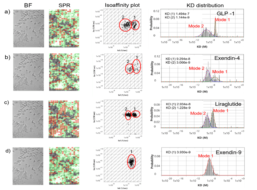
Figure 1: Peptide binding kinetics on GLP-1R overexpressed HEK293 cell surface.Bright field image of overexpressed HEK293 cells on the sensor surface and its corresponding SPR image. Red squares are regions of interest (ROIs) that observe responses which closely fit the kinetic binding model. The green regions indicate areas confluent with cells. Active areas designated by red ROIs overlap closely with cell regions indicating high cell specificity and low non-specific binding. The measured interactions of a) GLP-1, b) Exendin-4, c) Liraglutide, and d) Exendin-9 with GLP-1R receptors on the surface of HEK293 cells are presented in isoaffinity scatter plots to reveal binding heterogeneity and predominant modes of interaction. Two predominate binding modes (1 & 2) for the bivalent interaction are observed, showing similar on-rates but dissimilar off-rates. The KD histograms displaying mode (1) and mode (2) were extracted from each isoaffinity scatter plot and fitted with Gaussian distribution to statistically determine the mean and distribution of the kinetic parameter.
In this model, the C-terminus of the peptide first anchors to the extracellular domain (ECD) of GLP-1R (mode 1), facilitating alignment for a subsequent N-terminal insertion into the transmembrane domain (TMD) (mode 2). The two modes shared similar association rates but exhibited markedly different dissociation rates, a hallmark of bivalent interactions. Mode 1, attributed to ECD-only anchoring, represents a lower-affinity, faster-dissociating state, while mode 2, arising from dual ECD-TMD engagement, yields a higher-affinity, slower-dissociating complex. GLP-1 produced KD values of 1.1 nM and 149 nM for modes 2 and 1, respectively. Exendin-4 yielded KD values of 3 nM and 92 nM, while Liraglutide demonstrated the tightest overall binding with KD values of 1.2 nM and 29 nM. In contrast, the antagonist Exendin-9 produced a single Gaussian distribution in its KD histogram with a KD of 3.9 nM, consistent with its monovalent ECD-only binding mode. This single-mode behavior reflects the structural basis of antagonism: Exendin-9 lacks the N-terminal residues required for TMD engagement, thereby occupying the ECD binding site and blocking receptor activation without initiating downstream signaling. These kinetics-based observations are fully consistent with published biochemical, mutational, and structural studies of GLP-1R and other class B GPCRs.4
Among the agonists, Liraglutide exhibited the most favorable binding profile, with the narrowest distribution widths for both binding modes and the slowest dissociation rate. Narrow distribution widths reflect reduced conformational heterogeneity and more uniform receptor engagement, indicative of a stable, well-defined binding ensemble. This behavior is attributed to Liraglutide’s lipidation-mediated membrane association and optimized peptide architecture, which reduce conformational freedom and promote sustained dual-domain binding. These features are consistent with its superior in vivo pharmacology, including a prolonged plasma half-life of 11–15 hours compared to 1–2 minutes for native GLP-1, once-daily dosing regimen, and superior clinical outcomes demonstrated in head-to-head trials.5&6 GLP-1 showed the broadest distribution width in mode 2, indicative of greatest binding state heterogeneity in TMD engagement, suggesting that a subset of dual-domain binding events may not translate to productive receptor activation, a behavior consistent with partial agonism at the microstate level.
This study establishes SPRM as a powerful, label-free platform for dissecting the on-cell binding kinetics of GPCR–peptide interactions with single-cell resolution. By resolving dual binding modes arising from sequential ECD and TMD engagement, SPRM provides mechanistic insight into agonist activation efficiency and antagonist selectivity that is inaccessible to conventional receptor binding assays. The ability to quantitatively differentiate full and partial agonism based on kinetic heterogeneity, and to do so on intact cells in a native membrane environment, offers a compelling framework for early-stage drug discovery, particularly for the rational design and screening of next-generation GLP-1R-targeted therapeutics.
DOWNLOAD PDF
Download a PDF of Application Note 181: In Vitro Characterization of Agonist and Antagonist Peptide Binding Interaction Kinetics to GLP-1R in HEK293T Cells Using SPR Microscopy
- Li et al, Frontiers in Pharmacology 16 (2025): 1610512.
- Panjamaporn, et al, American Journal of Physiology-Endocrinology and Metabolism 327.5 (2024): E600-E615.
- Thirumurthy et al, ACS Medicinal Chemistry Letters (2026): 17 (5), 963–972.
- Wootten et al, Cell 165, no. 7 (2016): 1632-1643.
- Knudsen et al, Frontiers in Endocrinology 10 (2019): 155.
- Marso et al, New England Journal of Medicine 375, no. 4 (2016): 311-322.