
Plasmodium falciparum is responsible for most severe malaria cases, causing over half a million deaths annually. Central to erythrocyte invasion is the interaction between apical membrane antigen 1 (PfAMA1) and rhoptry neck protein 2 (PfRON2).1 The PfRON2(~4 kDa) ectodomain docks into a conserved hydrophobic cleft on PfAMA1 domain I (DI, ~30 kDa), and because no alternative invasion pathway exists, disrupting this interaction is an attractive antimalarial strategy. Chemical synthesis of PfAMA1 offers unique advantages over recombinant expression, including incorporation of non-natural amino acids and critical access to the D-enantiomeric mirror-image protein for biological display screening of D-peptide inhibitors.2
In this study, truncated 180-residue PfAMA1-DI construct (His123–Cys302) was chemically synthesized via a convergent six-segment native chemical ligation strategy. An AffiTag—comprising polyhistidine and biotin connected through a flexible 16-residue polyArg-containing spacer was appended to the cyclic construct to enable gentle Ni-NTA surface capture for SPR.3 SPR experiments were performed on the 5-channel BI-4500 instrument.
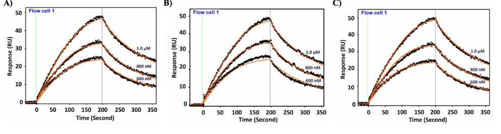
Figure 1: SPR sensorgrams from three independent replicates (A, B, C) of AffiTag-cyclicPfAMA1-DI binding to PfRON22021–2059 at 600 nM, 800 nM, and 1.0 µM. Solid orange lines represent 1:1 Langmuir fits to each concentration series. The average KD across the three replicates was 500.24 ± 14 nM.
Initial attempts to immobilize cyclicPfAMA1-DI directly on a dextran sensor chip via amine coupling, and separately to capture biotinylated PfRON2 on a streptavidin (SA) chip, both failed to yield reliable binding data — the former showing no interpretable signal and the latter exhibiting substantial non-specific binding of cyclicPfAMA1-DI to the streptavidin surface itself, likely due to disruption of the protein’s functional fold or surface-driven artefacts. These limitations motivated the switch to the gentler, orientation-controlled Ni-NTA/His-tag capture strategy. Folded AffiTag-cyclicPfAMA1-DI was captured on a nickel-NTA sensor chip via its polyhistidine tag; PfRON2 was passed as analyte at different concentrations (500 nM, 800 nM, and 1.0 µM). Binding was concentration-dependent and saturable (Figure 1). Global fitting to a 1:1 Langmuir model (Figure 2) yielded an average association rate constant ka = (1.42 ± 0.14) × 10⁵ M⁻¹s⁻¹, average dissociation rate constant kd = (7.08 ± 0.81) × 10⁻² s⁻¹, and average equilibrium dissociation constant KD = 500 ± 14 nM (Figure 1, Table 1).
| Measurement run | ka (M⁻¹s⁻¹) | kd (s⁻¹) | KD (nM) |
| A | 1.43 × 10⁵ | 6.95 × 10⁻² | 486.10 |
| B | 1.26 × 10⁵ | 6.34 × 10⁻² | 501.16 |
| C | 1.55 × 10⁵ | 7.95 × 10⁻² | 513.48 |
| Average | (1.42 ± 0.14) × 10⁵ | (7.08 ± 0.81) × 10⁻² | 500.24 ± 14 |
Table 1: Kinetic parameters for each of the three independent SPR replicates (A, B, C) of PfRON22021–2059 binding to AffiTag-cyclicPfAMA1-DI, with the resulting average ± SD (n = 3).
The entire binding experiment was independently repeated three times (Figure 1, panels A–C), and each replicate was fitted separately to a 1:1 Langmuir model to extract ka, kd, and KD (Table 1); the three independent estimates were then averaged to give the final reported values. Across the three replicates, ka ranged from 1.26×10⁵ to 1.55×10⁵ M⁻¹s⁻¹ and kd ranged from 6.34×10⁻² to 7.95×10⁻² s⁻¹, giving individual KD estimates of 486, 501, and 513 nM which supports the reproducibility of the measurement.
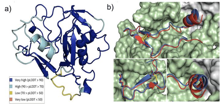
Figure 2: Structural comparison of the cyclic PfAMA1 Domain I (DI) construct and the PfAMA1 Domain I + Domain II (DI+DII) model. (a) Predicted structure of the cyclic PfAMA1-DI bound to PfRON2. (b) Overlay of the cyclic PfAMA1-DI model with the PfAMA1 DI+DII structure showing the absence of Domain II (gray) in the cyclic DI construct. Domain I is shown in green, Domain II in gray, and PfRON2 in red/blue.
PfAMA1-DI construct kinetic parameters were compared to the recombinant full-length PfAMA1-(DI+DII) (Figure 2). The comparison of kinetic parameters (Table 1) reveals that the ka of cyclic PfAMA1-DI is essentially identical to PfAMA1-(DI+DII) — 1.42 × 10⁵ vs. 1.49 × 10⁵ M⁻¹s⁻¹ respectively, demonstrating that encounter-complex formation is driven entirely by domain I and is independent of domain II. By contrast, the dissociation rate is ~22-fold faster in the truncated construct (7.08 × 10⁻² vs. 3.21 × 10⁻³ s⁻¹), accounting for the ~23-fold weaker (KD=22 nM) of 500 nM. This kinetic penalty is attributable to the absence of domain II, particularly the DII loop (Ala346–Lys395), which has been shown to prolong complex half-life 18-fold by kinetically locking the bound PfRON2 conformation through transient contacts with its N-terminal α-helix. Also, the KD of 500 nM also aligns closely with the affinity of a truncated PfRON2 variant lacking the N-terminal helix (~520 nM), providing independent structural corroboration.4
Despite the weaker absolute affinity relative to the full-length protein, cyclicPfAMA1-DI retains a functional PfRON2-binding groove and represents a viable scaffold for synthesis of the D-enantiomeric mirror-image protein, enabling mirror-image biological display campaigns to identify D-peptide or L-protein inhibitors of the PfAMA1–PfRON2 interaction. The Ni-NTA SPR format described here provides a reproducible, orientation-controlled kinetic assay for such drug discovery programs.
Materials is a summary of Mannuthodikayil et al, Chemistry–A European Journal 31, no. 28 (2025): e202500894
Author: Nguyen Ly, and Miyuki Thirumurthy | Biosensing Instrument | Published July 22nd, 2026
DOWNLOAD PDF
Download a PDF of Application Note 180: Surface Plasmon Resonance Analysis of Chemically Synthesized Cyclic Malaria Antigen PfAMA1 Domain I Binding to PfRON2
- Tonkin, M. L et al, Science 2011, 333 (6041), 463–467.
- Kent, S. B. H et al,Chem. Soc. Rev. 2009, 38 (2), 338–351.
- Mannuthodikayil et al, Chemistry–A European Journal 31, no. 28 (2025): e202500894
- Biswas et al, Biochemistry and Biophysics Reports 26 (2021): 100950.