Surface Plasmon Resonance Microscopy (SPRM) can directly resolve multiple kinetic binding modes in heterogeneous biological samples by extracting independent sets of kinetic parameters from spatially defined regions of interest (ROIs). Each ROI can be considered a separate measurement channel, from which association (ka), dissociation (kd), and equilibrium dissociation (KD) constants can be directly determined. When these independent sets of parameters are combined into affinity isotherms or histogram distributions to form a population-based analysis of the binding heterogeneity, multiple modes of binding interactions may be observed.
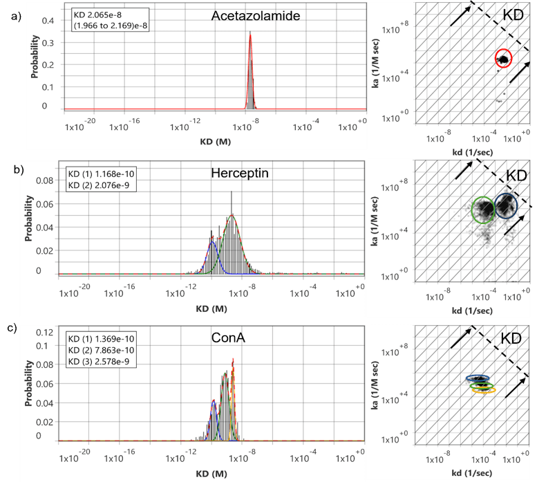
Figure 1: Heterogeneity of binding kinetics and affinity revealed by SPRM analysis. Left panels show distributions of equilibrium dissociation constants (KD) for three analytes: Acetazolamide (top), Herceptin (middle), and ConA (bottom). Right panels show the corresponding isoaffinity plots, where diagonal lines represent constant KD values (KD = kd/ka).
The simplest case is illustrated by small-molecule binding, where a single interaction mode dominates. Acetazolamide (223 Da) binding to Carbonic Anhydrase IX (CA IX) expressed on live suspension Ramos B cells produces a single, narrow KD distribution. Each ROI yields one consistent kinetic solution, and when aggregated, the histogram forms a single Gaussian peak (Figure 1a). This behavior reflects a uniform binding mechanism with minimal heterogeneity, as expected for a well-defined small molecule interaction with a single binding site.1
In contrast, more complex behavior emerges with multivalent interactions. Herceptin (trastuzumab), a bivalent antibody targeting HER2 on BxPC3 pancreatic cancer cells, produces two distinct peaks in the KD distribution. These two peaks arise from affinity and avidity contributions inherent to antibody binding. Importantly, this system is characterized by a single association rate constant (kₐ) but two distinct dissociation rate constants (kd), reflecting different dissociation pathways (Figure 1b). The higher-affinity (0.11 nM) population corresponds to bivalent engagement, where simultaneous binding of both antibody arms (2.08 nM) stabilizes the interaction and slows dissociation.2 The lower affinity population reflects one-arm binding resulting in rapid dissociation. In this case, the two-peak distribution is due to the intrinsic biophysical mechanism of the interaction specifically, the coexistence of affinity and avidity driven binding modes.
A fundamentally different scenario is observed with Concanavalin A (Con A) binding to BxPC3 cells, where multiple peaks arise from biological heterogeneity rather than multivalency (Figure 1c). 3 Unlike the Herceptin example, these peaks are not due to affinity versus avidity but instead represent distinct biological subpopulations within the sample. The multiple peaks therefore reflect differences in binding behavior across ROIs, arising from heterogeneity in glycan accessibility, density, and composition. In the case of Con A, three distinct modes of sugar residues are known to result in Con A binding.
| Compound | Target Receptor, Cell | # of Peaks | kₐ (M⁻¹ s⁻¹) | kd (s⁻¹) | KD (nM) |
| Small molecule – Acetazolamide | CA IX, on Ramos B Cell | 1 | 2.09 × 10⁵ | 4.36 × 10⁻3 | 20 |
| Bivalent antibody – Herceptin | HER2, on BxPC3 cell | 2 | 1.44 × 10⁶ 8.21 x 10⁶ | 2.51 × 10⁻⁴ 6.71 × 10⁻3 | 0.11 2.08 |
| Lectin – Con A | 1) Oligomannose-type N-glycans 2) Hybrid-type N-glycans 3) Complex type bi-antennary N-glycans on BxPC3 cell | 3 | 2.87 × 10⁵ 1.18 × 10⁵ 5.30 × 10⁴ | 4.50 × 10⁻⁵ 8.60 × 10⁻⁵ 1.30 × 10⁻⁴ | 0.13 0.78 2.60 |
Table 2: Summary of kinetic binding parameters for different ligand classes interacting with cell surface targets.
With the vast amount of high resolution and heterogeneous binding interaction data being collected by SPRM, predominant mechanisms of interaction can be readily identified using statistical tools. A single peak of interaction indicates that only a single predominant interaction mode is observed across all ROIs, as seen with small molecule Acetazolamide. However, two peaks with a shared association rate but distinct dissociation rates often indicates that avidity binding behavior is also present, as is demonstrated by the bivalent antibody Herceptin. Additionally, multiple peaks at various association and dissociation values can indicate both spatial (e.g. cell population-level variations) or compositional (e.g. receptor-level variations) in heterogeneity, as observed with Con A, which is a sugar binding lectin that is highly dependent upon variations in the post-translational cell environment.
Unlike traditional SPR, which produces a single aggregate binding response from the sensing area, SPRM leverages high resolution imaging to produce hundreds to thousands of localized and independent binding responses, thereby enabling detailed measurement of binding heterogeneity mapped across the entire sensing area. This unique ability enables differentiation between multiple mechanisms of binding interactions, making SPRM an invaluable tool for understanding complex interactions in heterogeneous biological systems.
Author: Nguyen Ly, Miyuki Thirumurthy, and Jesús Aguilar Díaz de león | Biosensing Instrument | Published June 10th, 2026
DOWNLOAD PDF
Download a PDF of Application Note 179: Resolving Multiple Kinetic Binding Modes in Complex Biological Systems
- Thirumurthy, M.A.et al ACS Medicinal Chemistry Letters, 17(1), pp.154-161.
- Aguilar Díaz de león, Jesús S et al Glycobiology 35, no. 12 (2025): cwaf066.
- Aguilar Díaz de león, Jesús S et al, Plos one 19, no. 5 (2024): e0304154.