Transmembrane carbonic anhydrase IX (CA IX) is a zinc metalloenzyme and overexpressed in numerous cancers and represents an attractive therapeutic target due to its limited expression in normal tissues and its key role in tumor pH regulation, metabolic adaptation, and drug resistance.1. Despite its importance, quantitative characterization of small molecule binding to CA IX remains difficult because purification of the full-length, membrane anchored enzyme often disrupts its native structure and microenvironment. As a result, many reported binding studies are performed using recombinant extracellular domains, lacking in native membrane association, which may misrepresent inhibitor access, binding kinetics, and affinity.2 Recent evidence has shown that CA IX is expressed in selected B-cell lymphomas, including Burkitt lymphoma, and there it plays a critical role in metabolic adaptation to hypoxia and cellular stress.3 Since hematologic cancer models are intrinsically nonadherent, investigation into CA IX in live suspension cells represents an important avenue toward the capture of physiologically relevant binding behavior.
SPRM provides single-cell spatial resolution and high sensitivity to binding induced dielectric changes, making it especially well-suited for investigating membrane associated targets on small hematopoietic cancer cells.4 The e work described here represents a pioneering effort in the demonstration of feasibility of SPRM for kinetic measurements of small molecule interactions on live suspension cells.
In the present study, SPRM was utilized for the kinetic characterization of interactions involving five
sulfonamide-based small-molecule inhibitors (Acetazolamide, Sulfanilamide, Furosemide, Dansylamide, and 4-Carboxybenzenesulfonamide (4-CBS)) with CA IX expressed on live Ramos B suspension cells.5 Sulfonamides are the most studied class of carbonic anhydrase inhibitors; they interact with the enzyme through coordination with the zinc ion in its catalytic site. Consequently, the proper characterization of sulfonamide binding in its native, membrane-bound state is particularly important for correct in vivo behavior prediction of these inhibitors.
SPRM measurements were in good agreement with previously reported affinity values and showed excellent reproducibility with a coefficient of variation of 6.8%. To further place these data in context, kinetic interactions with CA IX were compared to those of carbonic anhydrase II (CA II), a cytosolic isoform with a homologous active site that is highly stable outside its native environment and has been well characterized using conventional kinetic methods. Such direct comparison documented distinct kinetic profiles, highlighting how membrane anchoring and cellular context modulate CA IX binding dynamics in ways unattainable using isolated protein assays. Interestingly, sulfanilamide shows ∼16 times greater affinity for in vitro CA IX than for ex vitro CA II. While the catalytic domains of CA IX and CA II are largely homologous, the membrane-associated context of CA IX provides a more open polar pocket with favorable hydrogen bonding and electrostatic stabilization of the sulfanilamide which is absent when the receptor is extracted from its native membrane context. These findings strongly suggest that both the structural features of the active site and the membrane context may have worked together to enhance the binding of sulfanilamide to CA IX. The kinetic distributions observed in the case of furosemide reflect structurally heterogeneous binding due to its flexible molecular scaffold and various interaction geometries, leading to variable receptor engagement. These unique observations are not readily possible using traditional kinetic measurement methods, which require membrane disruption or labeling. (Table 1, Figure 1).
| Small molecules | Molecular weight (g/mol) | SPRM results for CA IX | Reported values CA II 40 | ||
| KD | ka (M-1 s-1) | kd (s-1) | |||
| Acetazolamide | 222 | 21 nM | 2.08E+05 | 4.68E-03 | 19 nM |
| Sulfanilamide | 172 | 311 nM | 4.40E+03 | 1.39E-03 | 5 µM |
| Furosemide | 330 | 491 nM | 1.10E+03 | 1.16E-03 | 513 nM |
| Dansylamide | 250 | 586 nM | 2.53E+03 | 2.34E-03 | 760 nM |
| 4-CBS | 201 | 870 nM | 1.10E+03 | 1.19E-03 | 893 nM |
Table 1: Kinetics of small molecule binding interactions to in vitro membrane-bound CA IX and ex vitro purified CA II influence of live native membrane on CA IX binding interaction.
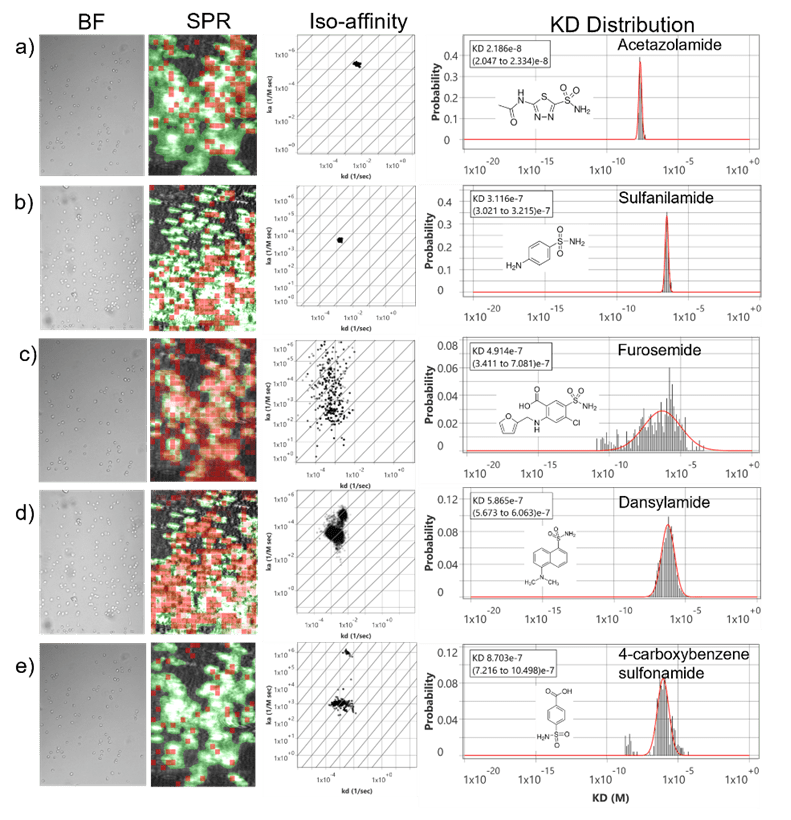
Figure 1. Small molecule binding kinetics on Live Ramos B cell surface. Bright field image of Ramos B cells on the sensor surface and its corresponding SPR image. Red squares are regions of interest (ROIs) that observe responses which closely fit the kinetic binding model. The green regions indicate areas confluent with cells. Active areas designated by red ROIs overlap closely with cell regions indicating high cell specificity. The measured interactions of a) Acetazolamide, b) Sulfanilamide, c) Furosemide, d) Dansylamide, and e) 4-CBS with CA IX receptors on the surface of live Ramos B cells are presented in iso affinity scatter plots to reveal binding heterogeneity and predominant modes of interaction. The KD histograms extracted from each iso-affinity scatter plot were fitted with Gaussian distribution to statistically extract the mean and distribution of the kinetic parameter.
In summary, this work positions SPRM as a powerful, label-free platform for quantifying small-molecule binding kinetics on live suspension cells in vitro. Maintaining the native receptor conformation and achieving single-cell resolution, SPRM supplies physiologically relevant information on membrane-associated drug targets otherwise poorly obtained by conventional approaches. These findings build a sound basis for extended use of SPRM in drug discovery and development, especially regarding membrane-bound targets in nonadherent cancer models, and open new avenues toward precise assessment of therapeutic inhibitors under conditions closely related to their in vivo environment.
Author: Nguyen Ly, and Miyuki Thirumurthy | Biosensing Instrument | Published January 27, 2026
DOWNLOAD PDF
Download a PDF of Application Note 173: Label-Free Kinetic Analysis of Carbonic Anhydrase IX Inhibitors on Live Suspension Cells Using Surface Plasmon Resonance Microscopy
- Pastorekova, S et al, Cancer and Metastasis Reviews. 2019, 38, 65.
- Hunter, S. A et al, In Methods Enzymol. 2016, 580.
- Chen, L. Q et al, Lymphoma 2015, 56 (5), 1432– 1439.
- Zhou, X. L et al, Angewandte Chemie - International Edition. 2020.
- Thirumurthy, Miyuki A et al, ACS Medicinal Chemistry Letters 2025