Surface Plasmon resonance microscopy (SPRM) enables high-resolution evaluation of the kinetic binding heterogeneity on whole single cells, without disrupting the native environment of the target. This capability permits measurement in unprecedented detail multiple binding modes such as affinity, avidity, and multivalent interactions.1 In this study, SPRM was employed to directly quantify the bivalent binding kinetics of three monospecific antibodies: Cetuximab (anti-HER1), Herceptin (anti-HER2), and anti-HER3 on whole BxPC3 pancreatic cancer cells. Kinetic interaction analysis for all three anti-HER antibodies was performed per SPRM cell and assay protocols. As can be seen from the isoaffinity plots and histograms, two predominant modes of interaction are consistently revealed for each antibody.
Occurrence of the two interaction modes is attributed to the bivalent structure of the antibodies, which is in reasonable contrast to the single interaction modes typically observed with monovalent interactions.2,3
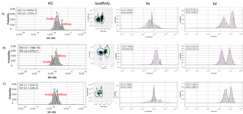
Figure 1: Antibody binding kinetics on native BxPC3 pancreatic cancer cells. Isoaffinity scatter plot extracted from thousands of responsive ROIs for (A) Cetuximab, (B) Herceptin and (C) anti-HER3 binding on native control BxPC3 cells. Two predominant binding modes (1 & 2) for the bivalent interaction are observed, showing similar on-rates but dissimilar off-rates. Mode (1) designates the double-arm avidity interaction and mode (2) designates the single-arm affinity interaction. KD histogram extracted from isoaffinity scatter plot taken along the diagonal axis kd/ka with Gaussian distribution fitted to analyze the kinetic interaction of all three antibodies on BxPC3 cells.
Quantitatively, Cetuximab showed KD values of 151 nM (affinity) and 4.6 nM (avidity), Herceptin exhibited 2.0 nM and 0.1 nM, while anti-HER3 showed 13 nM and 1.3 nM for the two respective binding modes (Table 1).
| Antibodies | Avidity KD (nM) | Affinity KD (nM) |
| Cetuximab | 4.6 | 151 |
| Herceptin | 0.1 | 2 |
| Anti-HER3 | 1.3 | 13 |
The presence of avidity interactions (mode 1) is supported by two key observations. First, both interaction modes exhibit comparable association rates (ka) but markedly different dissociation rates (kd), with the slower kd reflecting enhanced stabilization by the second binding arm (Figure 1 A-C).
Second, the dependence of the stronger binding mode on receptor density indicates that higher receptor abundance enhances the likelihood of bivalent binding, where the second antibody arm is more able to bind a neighboring receptor after the first arm binds (Figure 2).5&6 Therefore, the weaker monovalent interaction mode is attributed to single-arm affinity and the stronger bivalent interaction attributed to avidity.2
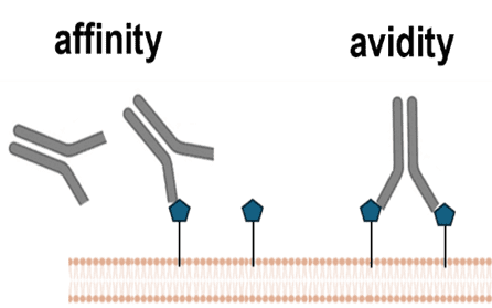
Figure 2: Schematic shows different avidity and affinity interaction scenarios.
The prevalence of such avidity interactions varied among antibodies as seen in Figure 3, comprising approximately 78% of total binding events for Cetuximab, 40% for Herceptin, and 58% for anti-her3, which corresponded with their relative receptor expression levels on BxPC3 cells.
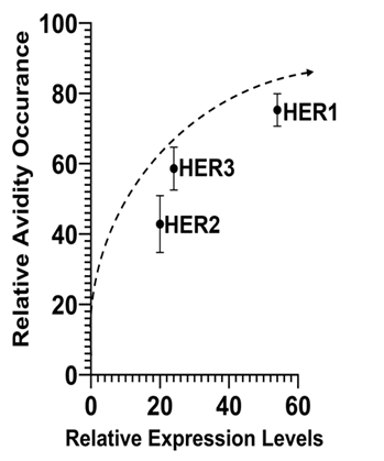
Figure 3: Avidity-receptor density dependence: Increasing relationship observed between the relative occurrence of avidity and receptor density. Relative expression levels obtained by fluorescence were plotted on the x-axis, and percent of total binding events observed for avidity were plotted on the y-axis.
This study demonstrates the unique ability of SPRM to resolve and quantify both affinity and avidity binding modes directly on whole, native cancer cells. The approach highlights how receptor density significantly impacts therapeutic antibody binding kinetics and provides key insights that can guide rational design and optimization of antibody-based therapies.
Author: Jesús Aguilar Díaz de León, Nguyen Ly, and Miyuki Thirumurthy | Biosensing Instrument | Published December 2nd, 2025
DOWNLOAD PDF
Download a PDF of Application Note 172: Affinity and Avidity Kinetic Binding Analysis Using SPRM
- Aguilar Díaz de león, Jesús et al, Plos One 19.5 (2024): e0304154.
- Miyuki Thirumurthy et al, Application Note 153, Biosensing Instrument (2025).
- Cruz, Victor L., et al, International Journal of Molecular Sciences 24.15 (2023): 12031.
- Marano, N et al, The Journal of Immunology, 143 (1989), 931–938.
- Erlendsson, Simon et al, Frontiers in Molecular Biosciences 7 (2021): 615565.
- Zhang, Fenni et al, Analytical Chemistry 87, no. 19 (2015): 9960-9965.