Traditional electrochemical (EC) techniques typically quantify averaged responses from a collection of individual elements and molecules on the surface of electrode.1 These approaches, while effective for bulk analysis, often overlook the complex heterogeneity inherent in many biological systems. This limitation underscores the imperative need for developing EC methodologies that can achieve spatially resolved detection. While scanning probe techniques offer greater spatial resolution, their temporal resolution is less optimal.2 On the contrary, optical methods deliver high temporal resolution with the trade-off of diminished spatial information. Within this spectrum of tools, EC-SPRM (surface plasmon resonance microscopy) emerges as a powerful technique, employing SPR to address both the temporal and spatial resolution and spatially resolve refractive index changes by electrochemical reactions in a label free manner.
EC-SPRM typically records localized changes on the electrode surface resulting from electrochemical processes. These plasmonic signals arise from redox reaction-induced refractive index variations near the metal film, alterations in the dielectric properties of the film (e.g., surface charge density), redox molecular binding events, or electrochemical deposition of additional layers onto the metal surface.3 In addition, the microscopy feature of EC-SPRM enables spatially resolved analysis of electrochemical activities on the electrode surface.4
This app note reviews two recently reported techniques for enhancing the sensitivity of EC-SPRM for electroactive biological systems: 1) “dye-sensitized” approach5 and 2) modification of the electrode surface with mesoporous silica film (MSF) to achieve sensitive imaging of the electroactive species.6
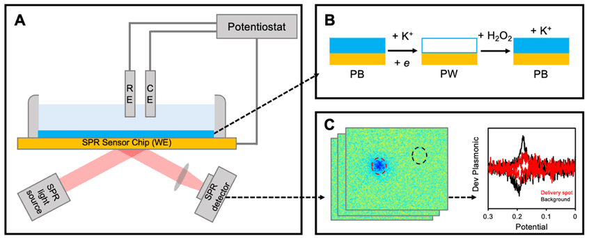
Figure 1. Schematic of the “dye-sensitized” approach (A), reaction mechanism at the Prussian blue (PB) film (B), and data visualization (C).
The first technique innovatively employs a redox-active dye coating on the SPRM sensor, significantly boosting sensitivity while allowing precise spatial resolution. The model application described here is the detection of hydrogen peroxide (H2O2), a reactive oxygen species with pivotal roles in various physiological processesand an established biomarker in health diagnostics. Conventional electrochemical assays—such as amperometry, voltammetry, and impedance spectroscopy—remain prevalent for H2O2 detection but often fall short in spatial resolution.7,8 This technique addresses these challenges by utilizing a Prussian blue (PB) nanofilm as a sensing layer on the sensing chip (Figure 1A), building on the earlier work demonstrating that Prussian blue nanoparticles (PBNPs) enable H2O2 detection via catalytic reduction.9,10
In the absence of H2O2, PB can be electrochemically reduced to Prussian white (PW), which generates a detectable increase in the SPRM electrochemical signal due to the refractive index change. In the presence of H2O2, some of the reduced form, PW, catalytically reduces H2O2 and converts back to PB (Figure 1B). This reduces the overall amount of conversion from PB to PW, causing a smaller refractive index change and a smaller increase in the EC-SPRM signal. As a result, the signal can be inversely correlated with H2O2 levels—a phenomenon that enables the establishment of a negative linear feedback relationship. This proportional relationship between the plasmonic signal and the concentration of H2O2 can then be used to map the local concentrations of H2O2(Figure 1C).
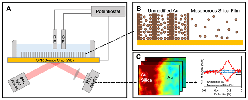
Figure 2. Schematic of the MSF approach (A), signal enhancement mechanism at the MSF (B), and data visualization (C).
To achieve spatially resolved sensing of heterogeneous systems with high sensitivity, the electrode surface can be modified. The second technique used by the researchers was to modify the gold (Au) electrode of the standard EC-SPRM setup with a mesoporous silica film (MSF) to achieve sensitive imaging of electroactive species (Figure 2A).6 Sensitivity enhancement occurs due to species nanoconfinement from the attraction of ions to the negatively charged silica films, thereby increasing the local concentration change and magnifying the signal (Figure 2B). The performance of Au-MSF electrodes in the setup was investigated using 1,1′-ferrocenedimethanol, carrying a positive charge in its oxidized form. There was a drastic enhancement of the sensing signal, with up to 37-fold improvement in the detection limit and up to 23 times improvement in the sensitivity (Figure 2C). Importantly, Au-MSF electrodes allowed for the quantification of detected concentrations, in contrast to Au electrodes. This type of nanoconfinement significantly broadens the potential applications of EC-SPRM, facilitating the plasmonic detection of various redox species at micromolar concentrations. Furthermore, Au-MSF electrodes also showed increased sensitivity for dopamine detection compared to Au electrodes and were able to visualize localized dopamine release.
These developments are promising for EC-SPRM based studies of biological systems, as they are capable of significant size selectivity, theoretically enabling exclusion of large entities (e.g., cell vesicles) while at the same time allowing smaller chemicals, such as neurotransmitters, to diffuse through. Furthermore, MSFs can also shield cells from the potentials applied during EC-SPRM experiments, reducing signal fluctuations due to cell movement and possible effects on cell metabolism. This research not only broadens the applications of EC-SPRM but also a step forward in the domain of electrochemical sensing, with significant promise for future bioanalytical applications.
This App note is based on the work from Prof. Yixian Wang’s research lab in the Department of Chemistry and Biochemistry, California State University, Los Angeles, Los Angeles, CA, USA.
DOWNLOAD PDF
Download a PDF of Application Note 165: Spatially Resolved Detection of Electroactive Biological Systems Using EC-SPR Microscopy
- Y. Wang, X. Shan, and N. Tao. Faraday discussions 193 (2016): 9-39. https://doi.org/10.1039/C6FD00180G
- A.G. Fallis, Royal Society of Chemistry, Cambridge, 2017 [Online].
- S. Wang, X. Huang, X. Shan, K.J. Foley, N. Tao, Anal. Chem. 82 (3) (2010) 935–941, https://doi.org/10.1021/ac902178f.
- Y. Wang, X. Shan, H. Wang, S. Wang, N. Tao, J. Am. Chem. Soc. 139 (4) (2017) 1376–1379, https://doi.org/10.1021/jacs.6b10693.
- A. Garcia, C. Dhoj, S. Groysman, K. Wang, S. Vo, A. Anguiano, T. Tran, D. Jiang and Y. Wang. Sensor Actuator Rep 8 (2024) 100218. https://doi.org/10.1016/j.snr.2024.100218
- S. Groysman, Y. Chen, A. Garcia, C. Martinez, K. Diego-Perez, M. Benavides, Y. Chen, Z. Wan, S. Wang, R. Liu, D. Wang, C. Liu, Y. Wang. ACS Electrochemistry (2025). https://doi.org/10.1021/acselectrochem.4c00227
- H. Sies, Redox Biol. 11 (2017) 613–619, https://doi. org/10.1016/j.redox.2016.12.035.
- M.A. O’Connell, J.R. Lewis, A.J. Wain, Chem. Commun. 51 (51) (2015) 10314–10317.
- Y. Zuo, Y. Jiao, C. Ma, C. Duan, Molecules 26 (11) (2021) 3352, https://doi. org/10.3390/molecules26113352.
- A. Garcia, K. Wang, F. Bedier, M. Benavides, Z. Wang, S. Wang, Y. Wang. Front. Chem. 9 (2021) 718666. https://doi.org/10.3389/fchem.2021.718666