Lysozyme (LZM) is found in body secretions like saliva, and it can catalytically hydrolyze peptidoglycans and exert catalysis-independent antimicrobial properties.1 Additionally, it is a valuable component in pharmaceutical and food products as it acts as a disinfectant, antihistamine and a preservative.1 Given its extensive use in both biological and industrial contexts, it is crucial to study the stability and the diverse biological interactions of different types of LZM in the presence of other proteins.2
Tannins (TA), found in numerous plant families, interact with proteins like LZM. The binding of tannins to such proteins primarily forms insoluble precipitates due to multiple hydrogen bonds formed between the phenolic hydroxyl groups of tannins and the carboxyl groups present in various proteins (the astringency mechanism).3&4 Biopharmaceutical formulations have been designed to use this astringency mechanism for the sustained-release system for LZM, by mixing the enzyme with TA, known as the tannylation process.5 The above mentioned sustained release system can achieve prolonged therapeutic effect by gradually releasing LZM over an extended period of time following the administration. However, only a limited number of studies have focussed on the LZM-TA interactions within the realms of food science and oral biology.
In this study, the nature of the interaction between tannic acid and lysozyme was studied using surface plasmon resonance (SPR) for the first time.6 SPR is an optical phenomenon that allows real-time and label-free quantification of molecular interactions with high sensitivity and specificity, requiring a small volume of sample for analysis. SPR has gained popularity for the in-vitro assessment of molecular interactions between ligand and analyte, providing valuable biophysical data such as kinetics, affinity, and thermodynamics.7
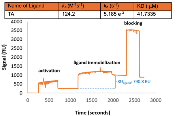
Figure 1: SPR sensorgram of CEWLZM immobilization step on functionalized gold surface with the kinetic values for the CEWLZM-TA interaction.
For the SPR experiments, chicken egg white lysozyme (CEWLZM) was chosen as a model protein.8 The measurements were performed after the immobilization of CEWLZM on the gold sensor surface using self-assembled long chain alkanethiol monolayer (Figure 1).9 This was followed by serial injections of different concentrations of TA to obtain the association (ka), dissociation (kd) rate constants, and equilibrium constant (KD) from the kinetics of the binding curves.
According to the results, ka, kd, and KD values were determined to be 124.2 M−1 s−1, 0.0051 s−1, and 41.7 µM, respectively. Also, molecular docking studies and molecular dynamics simulations were conducted to understand the binding mechanism of CEWLZM to TA and other inhibitors like Tri-N-acetylchitotriose (NAG3). In the SPR binding analysis supported by in silico studies, the KD value of CEWLZM-TA was closely aligned with the reported CEWLZM-NAG3 complex affinity value, such as 39.8 ± 8.8 µM. The KD for the CEWLZM-TA interaction also closely matched the KD value for the CEWLZM interaction with similar exogenous compounds like published in the literature indicating the presence of a relatively high affinity between CEWLZM and TA.6
SPR serves as a versatile and reliable platform for real-time monitoring of protein–compound binding, facilitating the determination of affinity constants in these ligand–analyte interactions. Importantly, the KD value of CEWLZM-TA is consistent with in silico investigations and supports studies conducted by different bio-interaction analysis techniques. This study highlights the efficacy of SPR as a valuable real-time monitoring tool for binding interaction analysis.
Author: Nguyen Ly and Miyuki Thirumurthy | Biosensing Instrument | Published January 21st, 2025
DOWNLOAD PDF
Download a PDF of Application Note 161: Surface Plasmon Resonance Identifies Lysozyme Interaction with Tannic Acid
- Liburdi, K., Benucci, I., & Esti, M. (2014). Comprehensive Reviews in Food Science and Food Safety, 13(5), 1062–1073
- Quan, T. H., Benjakul, S., Sae-leaw, T., Balange, A. K., & Maqsood, S. (2019). Trends in Food Science and Technology, 91, 507–517.
- Khanbabaee, K., & van Ree, T. (2001). Natural Product Reports, 18(6), 641–649.
- Khan, N. S., Ahmad, A., & Hadi, S. M. (2000). Chemico-Biological Interactions, 125(3), 177–189.
- Utatsu, K., Motoyama, K., Nakamura, T., Onodera, R., & Higashi, T. (2023). International Journal of Pharmaceutics, 643(June), 123229.
- Türkoğlu, E. A., Taştekil, I., & Özbek Sarica, P. (2024). Food Science & Nutrition, 12(10), 7392–7404.
- Gade, A., Sharma, A., Srivastava, N., & Flora, S. J. S. (2022). Clinica Chimica Acta, 527(January), 79–88.
- Jalili-Firoozinezhad, S., Filippi, M., Mohabatpour, F., Letourneur, D., & Scherberich, A. (2020). Materials Today, 40, 193–214.
- Su, X., Wu, Y. J., Robelek, R., & Knoll, W. (2005). Frontiers in Bioscience, 10(1), 268–274.