Alzheimer’s disease (AD) is currently ranked as the sixth leading cause of death in the United States and is the most common neurological disorder (1). Production and accumulation of amyloid-beta (Aβ) peptides in the brain are a hallmark of AD. Among Aβ peptides of different lengths, Αβ(1-42) has the highest propensity to aggregate (2).
Natural compounds and synthetic molecules have been screened as small-molecule drugs for disrupting the Αβ aggregation. A number of natural compounds from fruits, vegetables, teas or traditional herbal medicines have been shown to abolish Αβ-induced neurotoxicity on neurons (3). In this study, the binding interactions of two natural compounds with Αβ(1-42) were measured.
Ginnalin A (GA), a phenolic compound isolated from the red maple (Acer rubrum) species, has been reported to possess anticancer, antiglycan and antioxidative properties. GA is a 468.4 Da molecule and was identified to bind strongly with Aβ(1−42) monomers. Thus, GA is a potential drug candidate capable of delaying the onset of AD by reducing the number of oligomeric aggregates (3). The second compound, tabersonine (336.4 Da), is an indole alkaloid extracted from the beans of Voacanga. It is also an inhibitor of Aβ(1−42) aggregation. As a traditional medicine in Africa, Voacanga has been used to treat a wide range of diseases, including leprosy, convulsions in children, and infant tonic seizures. The decoction of the stem bark and root can also effectively treat mental disorders and be used as the precursor of vincristine for cancer chemotherapy (4).
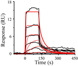
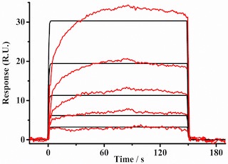
A research group in China used BI-4000 SPR to study the interactions between Aβ(1−42) monomers and GA as well as with tabersonine. Aβ(1−42) monomers were covalently linked to activated carboxyl groups on carboxymethylated dextran chips. For the GA experiment, the running buffer was 20 mM PBS, which was used to prepare GA solutions. Different GA concentrations (1.0, 2.3, 7.0, 22.0, 67.0, and 200.0 μM) were delivered to the detection and reference channels. The sensorgrams (Figure 1) were simulated using the 1:1 Langmuir isotherm binding model. Kinetic analysis yielded a dissociation constant (KD) of 3.5 ± 1.1 μM, kon of 0.49 ± 0.22 μM-1s-1, and koff of 0.015 ± 0.005 s-1. For the tabersonine binding assay, 100 mM PBS containing 100 mM NaCl was used as the running buffer. Tabersonine solutions of 200, 100, 50, 25, and 12.5 μM were injected to the detection and reference channels, yielding 535 ± 66 μM as KD (Figure 2).
GA prolongs the lag phase of Aβ(1−42) fibrillogenesis, while tabersonine does not. This explains why KD of GA is two orders of magnitude stronger than that of tabersonine (3). Moreover, the 1:1 binding model fits GA/Aβ(1−42) sensorgrams much better. Indeed, results of molecular dynamics simulations confirmed that GA interacts with Aβ(1−42) monomers in a single mode, whereas there are multiple interaction sites between tabersonine and Aβ(1−42). These two studies demonstrate that SPR is an effective and facile method for screening natural compounds as potential therapeutic drugs for neurological disorders and for gaining insight into the binding modes.
DOWNLOAD PDF
Download a PDF of Application Note 137: Ginnalin A and Tabersonine Inhibition of Amyloid β(1-42) Aggregation
- https://www.nia.nih.gov/health/alzheimers-disease-fact-sheet
- Stakos, D et al, J Am Coll Cardiol. 2020 Mar, 75 (8) 952-967
- Fan, Q et al, ACS Chem. Neurosci. 2020, 11, 4, 638–647
- Kai, T et al, ACS Chem. Neurosci. 2015, 6, 879−888