HER2 (Human Epidermal Growth Factor Receptor 2) is an important therapeutic target in different types of cancer because it functions to advance tumor development and growth.1 Antibody-based drugs such as Trastuzumab (Herceptin) have been extremely effective at treating HER2-positive tumors, so it is important to be able to determine the functional presence and availability of the receptor on cell types.2 In this study, Surface Plasmon Resonance Microscopy (SPRM) based on a two-well chip was applied to investigate the binding kinetics of Herceptin to HER2 receptors on two cell lines: BxPC3 pancreatic cancer cells and JIMT-1 Herceptin resistant breast cancer cells.
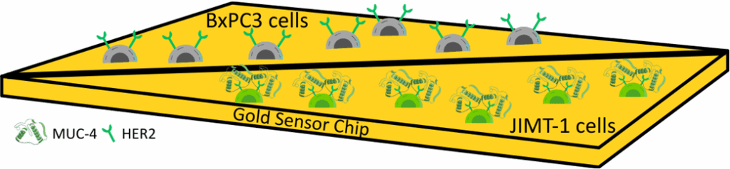
Figure 1: Schematic showing the SPRM setup with the two-well SPRM chip which has BxPC3 cells on one side and JIMT-1 cells seeded on the other side of the barrier.
SPRM is a powerful, label-free, and real-time analytical method for studying molecular interactions on the intact cell surface directly. The absence of spatial resolution in conventional methods like SPR, SPRM detects the binding events at the subcellular level, revealing receptor expression and heterogeneity.3 The use of a two-well SPRM chip enhances this capability even further by allowing two cell types to be measured in a single experiment, thereby improving efficiency and drastically minimizing variation.
In the current study, BxPC3 cells, with whom HER2 expression and responsiveness to Herceptin have been previously confirmed, were combined with JIMT-1 cells, which, as previously reported, lack functional binding response despite HER2 expression due to the masking of the HER2 receptor by a glycoprotein named MUC-4.4 This setup allowed for side-by-side real-time observation of specific versus non-specific interactions (Figure 1).
Regions of interest (ROIs) were defined around individual cells from each population for analysis of the binding interaction. The same number of ROIs was selected for both cell types to enable equal comparison. When Herceptin was injected, SPRM quantified the binding response and generated two data files for the two cell lines, measuring both binding signal and kinetic values. The red squares on the binding activity map reveal high amounts of cell-specific binding activity for Herceptin on BxPC3 cells and the kinetic analysis revealed two distinct modes of binding of Herceptin to HER2 on BxPC3 cells, consistent with the antibody bivalency and heterogeneity of receptor presentation. Mode 1 exhibited high affinity binding with rapid association and slow dissociation, while Mode 2 indicated lower affinity interaction due to likely steric constraint or adverse receptor orientation (Figure 2B).
In contrast, there was no response to binding observed in the JIMT-1 cells, mirroring the lack of functional HER2 interaction (Figure 2C). This unbinding is an essential control, guaranteeing the specificity of Herceptin binding to HER2 on target cells and highlighting the significance of the use of a functional negative cell line in these studies. Comparing two biologically relevant models in parallel under the same conditions ensures the practicality of the two-well SPRM chip not only to reduce time and cost but also to enhance the confidence of the data.

Figure 2: Herceptin interaction with BxPC3 and JIMT-1 cells. A) Bright field image of a two-well SPRm chip with the HER2 positive – BxPC3 cells on the top and functional negative HER2 -JIMT-1 cells on the bottom divided by a barrier blank area. B) Histograms describing total kinetic interactions and distributions for Herceptin on BxPC3 cells C) Histograms describing the lack of kinetic interactions for Herceptin on JIMT-1 cells.
In conclusion, this report demonstrates the application of SPRM with a two-well sensor chip which includes a functional negative control, enabling more accurate kinetic profiling for characterizing therapeutic mechanisms and resistance in cancer biology.
Author: Nguyen Ly, Miyuki Thirumurthy, and Jesús Aguilar Díaz de león | Biosensing Instrument | Published September 17th, 2025
DOWNLOAD PDF
Download a PDF of Application Note 170: Simultaneous Kinetic Analysis of HER2 Binding in Pancreatic and JIMT-1 Cancer Cells Using SPRm 220
- Tai et al., Journal of controlled release 146, no. 3 (2010): 264-275.
- Bradley et al, The Lancet Oncology, 22(8), (2021) pp.1139-1150.
- Aguilar Díaz de león, Jesús et al ,Plos one 19, no. 5 (2024): e0304154.
- Ghasemi et al, Oncogenesis, 3(8),(2014) pp.e117-e117.